 |
||
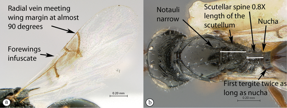
| 1 | Forewings infuscate over area surrounding venation (a). Marginal cell 1.55× as long as wide (a), venation thick with a very thin marginal vein (a); radial vein meeting wing margin at almost 90 degrees (a). Scutellar spine long, 0.8× length of the scutellum (excluding spine) (b). Notauli narrow (maximum width 0.35× the minimum distance separating notauli towards posterior mesoscutal margin) (b). Head subquadrate, 1.1× wider than long. First tergite (petiole) long (0.6× as long as high in lateral view; twice as long as nucha in dorsal view) (b) | Xyalophora tedjoansi sp. n. |
 |
||
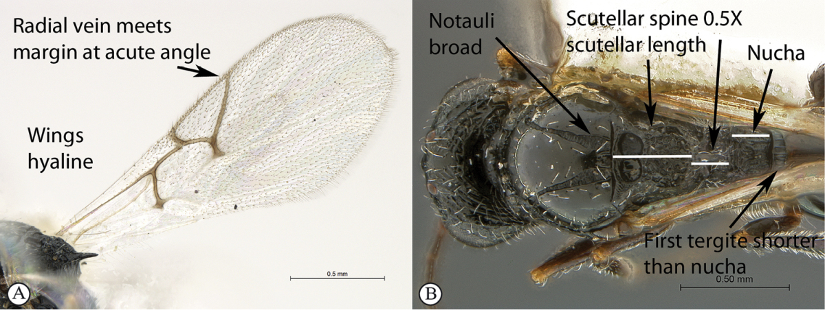
| – | Forewings hyaline (A). Marginal cell 2–2.5× as long as wide (A), venation thinner with less contrast in thickness with marginal vein (A); radial vein meets wing margin at acute angle (A). Scutellar spine shorter, 0.5× the length of the scutellum (excluding spine) (B). Notauli widened posteriorly (maximum width 0.7–0.8× the minimum distance separating notauli towards posterior mesoscutal margin) (B). Head distinctly (1.25×) wider than long. First tergite short (0.2–0.25× as long as high in lateral view; either a third of nucha length (B) or equivalent in length to nucha in dorsal view) | 2 |
 |
||
| 2 | Marginal cell 2.5× as long as wide (a). Nucha short, equivalent in length to first tergite (b). First tergite 0.25× as long as high in lateral view (b). Second flagellar segment longer than first. Median mesoscutal impression small (c) | Xyalophora provancheri Jiménez & Pujade-Villar |
 |
||
| – | Marginal cell twice as long as wide (A). Nucha long, 3× first tergite length (B). First tergite 0.2× as long as high in lateral view. Second flagellar segment as long as first. Median mesoscutal impression large, distinct (C) | Xyalophora tintini sp. n. |

An official website of the United States government
Here's how you know
Official websites use .gov
A
.gov website belongs to an official
government organization in the United States.
Secure .gov websites use HTTPS
A lock (
) or https:// means you've safely
connected to the .gov website. Share sensitive
information only on official, secure websites.