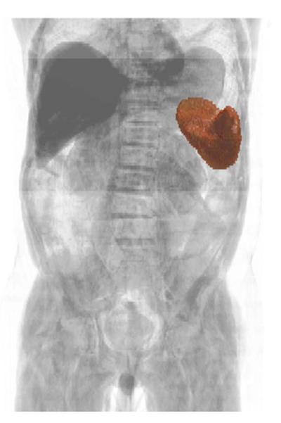
Fig. 3.
Volume-rendered CT of the abdomen and pelvis with overlaid 3-D surface rendering of the spleen, segmented by a fully automated multi-atlas content labeling algorithm. This technology is under investigation as a means of efficiently and accurately extracting spleen volume data for biomarker analyses. (Image courtesy of Zhoubing Xu, Ph.D. graduate student, Vanderbilt University.)

