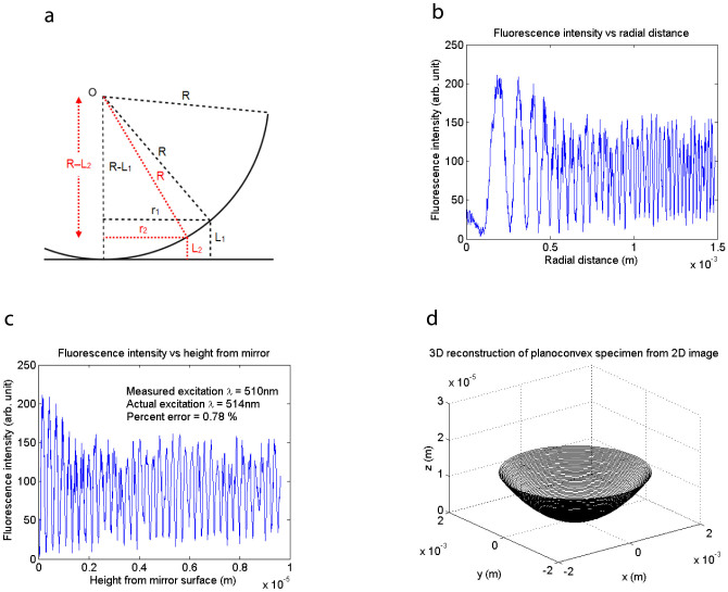
Figure 2. Three-dimensional reconstruction from a single two-dimensional image.
(a) Diagram of planoconvex specimen in contact with the mirror. The height L1 − L2 separating two successive bright fringes of radii r1 and r2 is given by  . (b) Fluorescence intensity in Fig. 1c plotted versus radial distance, showing the fringe spacing getting smaller with distance from the centre, consistent with a fluorescent shell of spherical shape excited by the evenly-spaced antinodes of a standing wave. (c) Fluorescence intensity plotted versus height from the mirror surface, obtained using the geometry in (a), showing evenly-spaced peaks where the dye cut the antinodes of the standing wave. The measured antinodal spacing is 255 nm, accurate to within 0.78% of the actual value of λ/2n = 257 nm using an excitation wavelength of 514 nm and a refractive index of n = 1 (air). (d) Three-dimensional reconstruction of planoconvex specimen from the two-dimensional fluorescence image in Fig. 1c.

