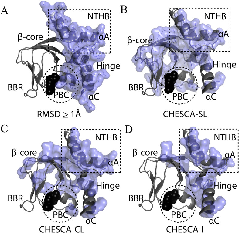
Figure 6. Summary maps of CHESCA-based allosteric ensembles identified for PKA RIα CBD-A.
(A) Structure-based allosteric residues for the folded CBD-A. Residues with a local RMSD between the C-bound and cAMP-bound crystal structures of PKA R (PDB IDs: 3FHI and 3PNA) greater than 1 Å are shown as a blue surface. (B–D) Allosteric residues from the CHESCA-SL, -CL and -I analyses of PKA CBD-A, respectively, mapped onto its crystal structure. cAMP is shown as black spheres.

