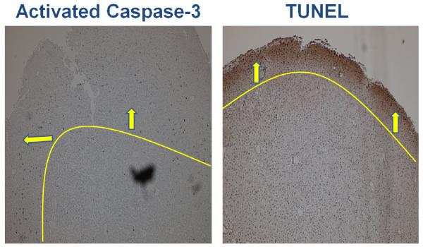
Figure 3. Distribution of Injury Following In Vitro Rediffusion.
Following 6 hours of rediffusion, there is a peripheral pattern of apoptosis. Both activated caspase-3 and TUNEL stain with greater intensity toward the periphery of liver cubes subjected to IR. This pattern of rediffusion injury is in contrast to the centralization of ischemic injury. Lines / arrows have been superimposed to demarcate the peripheral pattern of injury. Images are at 4x magnification.

