
Figure 3.
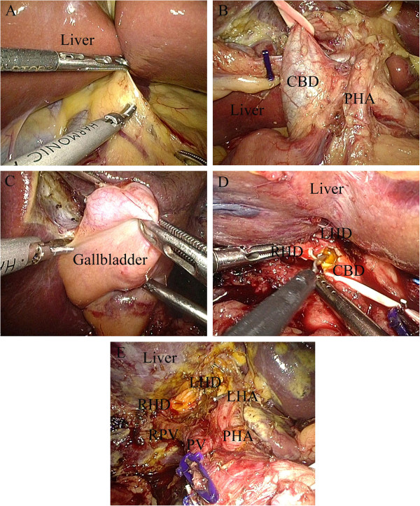
Resection of mass on biliary duct. (A) Division of hepatoduodenal ligament. (B) Visualization of properhepatic artery, common bile duct, and portal vein. (C) The cystic artery was clipped and divided while the cystic duct was clipped and left in situ. (D) Transection of left and right hepatic ducts 1.0 cm above the mass. (E) Traversal of common bile duct about 0.5 cm below the mass. CBD, common bile duct; LHA, left hepatic artery; LHD, left hepatic duct; PHA, proper hepatic artery; PV, portal vein; RHD, right hepatic duct; RPV, right portal vein.