
Figure 1.
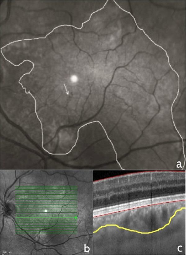
Near infrared reflectance and enhanced depth imaging optical coherence tomography cross-section of retinal and choroidal alterations in NF1. Near infrared reflectance image of “corkscrew” or “spiral” microvascular alteration (arrow) overlying large patchy choroidal alteration delineated with a white line (a), raster optical coherence tomography image and corresponding cross-section of the choroidal nodule delineated with a yellow line (b and c).