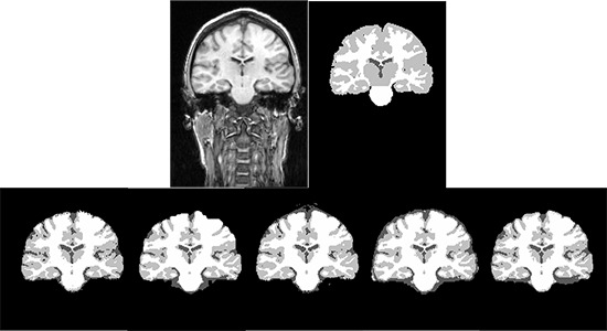
Figure 3.

The segmented GM, WM and CSF from a selected subject from IBSR real MR images. The first row shows the input MR image and the ground truth for GM, WM and CSF. The second row from left to right shows the segmentation results using SPM8-Seg, SPM8-VBM, SPM8-NewSeg, FSL and Brainsuite, respectively.
