Figure 3.

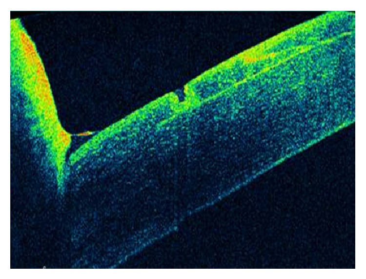
In the lower quadrant, the cut done primarily with the microkeratome opened up from inside after colliding with the new femtosecond flap, leading to an epithelial defect that was visualized with the OCT system.

In the lower quadrant, the cut done primarily with the microkeratome opened up from inside after colliding with the new femtosecond flap, leading to an epithelial defect that was visualized with the OCT system.