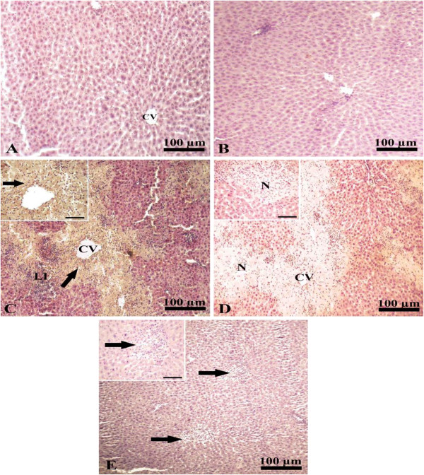
Figure 2.

Immunohistochemical staining of MMP-8 in liver. A and B, liver of CTR and CUR administered groups immunostained showing normal hepatic architecture with presence of a central vein (cv) and normal hepatic cords. C, liver of paracetamol intoxicated group showing increased expression of mmp-8 (arrow) in the necrotic area surrounding central vein (cv) together with leukocytic infiltration (neutrophils and lymphocytes; LI). D, liver of control negative paracetamol intoxicated group with no expression of mmp-8 in the necrotic area (N) around central vein (CV). E, liver of paracetamol intoxicated group treated with curcumin immunostained showing no expression of mmp-8 (arrows). Scale bar for photos from A to E is 100 μm. Inserts are high magnification fields in C, D and E with scale bars of 50 μm.
