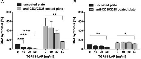
Figure 3.

Attenuated proliferative response of bead-silenced T cells to TCR/CD28-activating culture dishes. T cells were incubated in round-bottom tubes with anti-CD3/CD28/LAP beads which were loaded with indicated amounts of TGFβ1-LAP. After A) 15 min and B) 24 h bead/T cell activation reactions were transferred onto tissue culture plates which were either left untreated (black bars) or were coated with anti-CD3 and anti-CD28 antibodies (grey bars) *, **, *** depict significant (p <0.05, 0.01, 0.005) attenuation of proliferation rates when T cells were preincubated with TGFβ1-LAP on activating beads. A) Transfer of 15 min-incubated bead/T cell conjugates to uncoated plates recapitulated dose-dependent attenuation of T cell proliferation by TGFβ1-LAP on activating beads. These T cells strongly responded to independent anti-CD3/CD28 stimulus on culture plates. This additional proliferative response was inhibited by initial TGFβ1-LAP-presentation in dose-dependent manner; down to ~31% at highest loading of TGFβ1-LAP (n = 4). B) After 24 h of bead/T cell preincubation T cells responded with additional proliferation to plate-bound anti-CD3/CD28 antibodies. Proliferation on anti-CD3/CD28 plates, however, was restored to nearly the same rates with and without TGFβ1-LAP preincubation on beads; down to ~85% at highest doses (n = 3).
