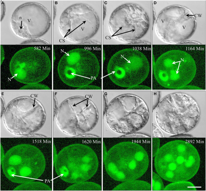
Figure 2.
Time-lapse of type I development (embryogenic pollen) shown by synchronously acquired DIC and fluorescence images. (A) Uni-nucleate pollen (microspore) with nucleus close to pollen aperture. (B,C) The nucleus has migrated away from the pollen periphery and cytoplasmic strands are formed. The blurred fluorescence signal indicates the break-down of the nuclear envelope prior to mitosis. (D) Newly formed cell wall (DIC) and a pair of daughter nuclei (GFP) after mitosis. (E) Appearance of cytoplasmic strands indicating imminent second mitosis. (F,G) Newly formed intermediate cell wall (DIC) separates four cells (GFP) contained within the pollen envelope. (H) Additional cycles of mitosis create a multicellular structure. CS, cytoplasmic strand; CW, cell wall; N, nucleus; n, nucleolus; PA, pollen aperture. Bar = 20 μm.

