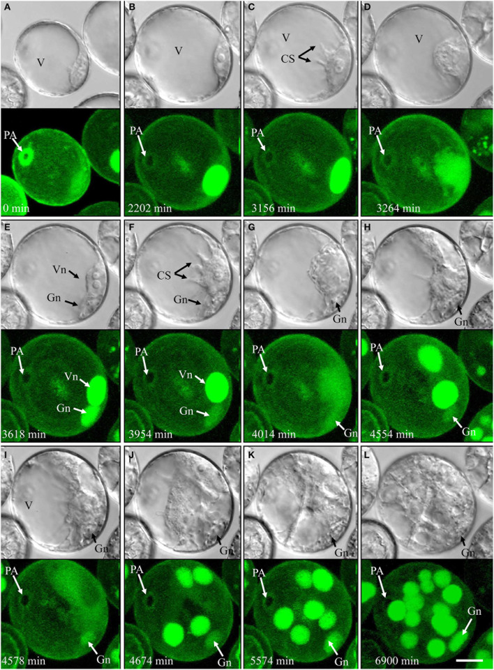
Figure 4.
Time-lapse of type IV development (embryogenic pollen) shown by synchronously acquired DIC and fluorescence images. (A) Uni-nucleate pollen with large vacuole and thin layer of peripheral cytoplasm. (B) Uni-nucleate pollen increases in size. (C) Cytoplasmic strands appear prior to pollen mitosis I. (D–F) Large spherical vegetative-like nucleus and smaller ellipsoid generative-like nucleus after asymmetric division. (G–L) Synchronized mitotic events originate from the vegetative-like cell; note that the generative-like cell does not show any mitotic activity. CS, cytoplasmic strand; Gn, generative-like nucleus; PA, pollen aperture; V, vacuole; Vn, vegetative-like nucleus. Bar = 20 μm.

