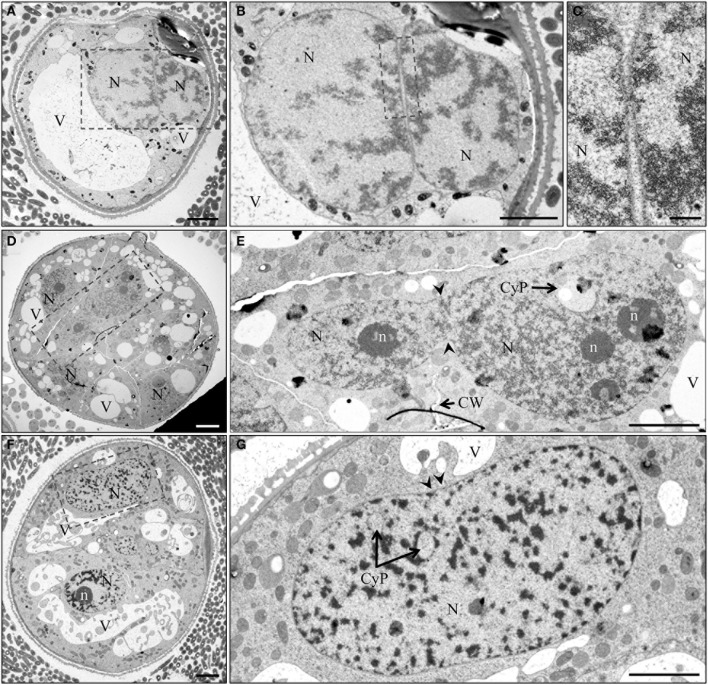
Figure 7.
TEM micrographs of nuclear fusion at different stages of pollen embryogenesis. (A) Induced immature pollen during first day of culture with two nuclei in close vicinity. (B,C) Detail of (A) shows the absence of cell wall and the close proximity of the nuclear envelopes. Arrow indicates region of assumed membrane fusion. (D) Multicellular structure 7 days after initiation of pollen embryogenesis. (E) Two nuclei after fusion with incomplete cell wall formation near the site of nuclear fusion. (F) Multicellular structure 7 days after initiation of pollen embryogenesis. (G) Elongated nucleus with clear median constriction (arrowheads), cytoplasmic pockets and a narrow median band marking the site of fusion. CyP, cytoplasmic pocket; CW, cell wall; N, nucleus; n, nucleolus; V, vacuole. Bar = 20 μm.

