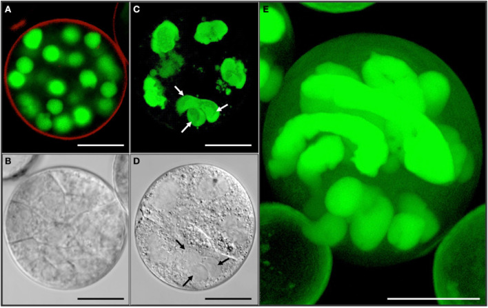
Figure 8.
Variable ploidy level in a multicellular structure shown by synchronously acquired DIC and fluorescence images. (A,C,E) GFP. (B,D) DIC. (A,B) Haploid multicellular structure with cell walls and spherical nuclei. (C,D) Chimeric polyploid multicellular structure with irregular shaped nuclei often not separated by cell wall. Note the difference of nuclear sizes. Arrows refer to a possible triple fusion. (E) Multicellular structure with highly polyploid nuclei next to small spherical, likely haploid, nuclei. Bar = A,B,D–E = 10 μm; C = 1 μm.

