
Figure 2. Preclinical staging of Alzheimer’s disease and short-term progression rates.
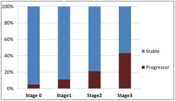
Using the preclinical staging criteria, at fixed cut-points corresponding to 90% sensitivity for diagnosing AD and the 10th percentile of cognitive scores of cognitively normal individuals, Stage 0 corresponds to low Aβ load on PET and absence of imaging markers of neuronal injury (i.e. normal hippocampal volumes on MRI and/or absence of AD-like pattern of hypometabolism on PET); Stage 1 corresponds to high Aβ load on PET and absence of imaging markers of neuronal injury; Stage 2 corresponds to high Aβ load on PET and presence of imaging markers of neuronal injury; Stage 3 corresponds to low Aβ load on PET, presence of imaging markers of neuronal injury, and subtle cognitive impairment. The proportion (%) of patients who progressed to mild cognitive impairment during a median follow-up of 15 months is demonstrated. Diagnosis of MCI was made according to Petersen criteria, 90 blinded to the imaging biomarker data used for staging.