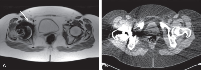
Figure 1.
Patient 6. Type-2a lesion (indicated by arrow) classified on MARS MRI scan (A) but lesion cannot be seen on the equivalent CT scan (B). The high attenuation coefficient of the metal implant on CT has led to significant scatter obscuring much of the periprosthetic anatomy, further compounded by a less clear distinction of soft tissues with this modality.

