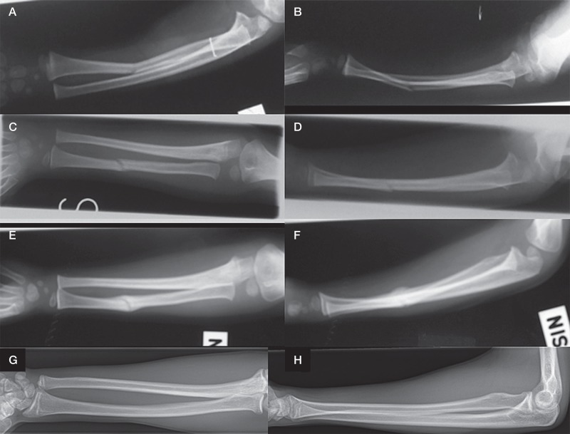
Figure 1.
An illustrative series of radiographs taken from a case who participated in the study. A 4-year-old boy suffered from a left-side both-bone forearm shaft fracture in the middle third. A. and B. There was a greenstick fracture in the radius and plastic bowing in the ulnar shaft. C. and D. 3 weeks after closed reduction and cast immobilization, the forearm presented good alignment in 2 directions. Slight malalignment remained in the bowed ulna. E. and F. 6 weeks after the injury, free mobilization was allowed. However, worsening alignment with both posterior and radial angular curvatures in the radius was seen (panel F). G. and H. Long-term radiographs 11 years after the injury show good alignment without any other bone complication. The remaining lateral bowing of the radius does not exceed 15° and is consistent with anatomic variation (panel G).

