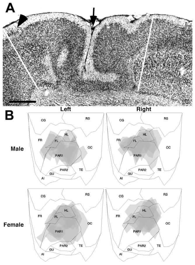
Fig. 2.
(A) Photomicrograph of a typical neocortical malformation following freezing injury to the developing cortical plate. In comparison to the normal six-layered cortex (right), the malformation consists of a sulcus (arrow), and four layers. Layer i is contiguous with the molecular layer. Layer ii is contiguous with layers II–III of the intact neocortex. Layer iii is the lamina dissecans, and consists of the remnants of the cortical plate that is damaged by the freezing injury. Layer iv, when present, is contiguous with the subplate (layer VIb). Arrowhead indicates a layer ii dysplasia. Scale bar=500 μm. The lower half of the figure is flattened maps of the neocortex illustrating regions of damage of the left and right hemisphere of male and female rats used in the experiment 1.

