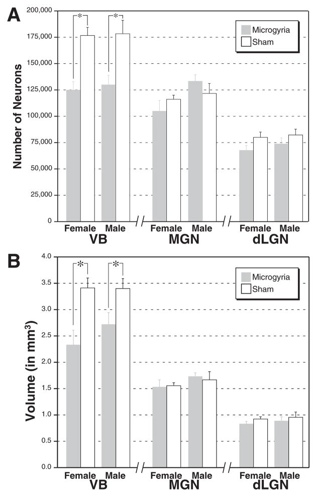
Fig. 3.
(A) Mean (±S.E.M.) number of neurons in the VB, MGN, and dLGN nuclei of the thalamus in male and female rats either with microgyria (dark bars) or shams (white bars). In VB, both male and female subjects with microgyria have significantly fewer neurons than their sham counterparts. There are no significant differences in neuron number in other thalamic nuclei. (B) Mean (±S.E.M.) volume of thalamic nuclei in subjects with and without induced malformations of the somatosensory neocortex. The volume of VB is significantly decreased in both male and female subjects with microgyria (dark bars) when compared with shams (white bars). There are no significant differences in volume in MGN or dLGN.

