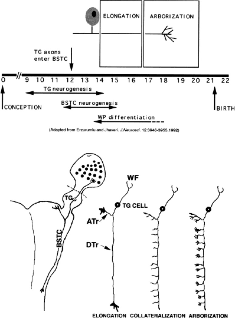
Fig. 1.
Developmental history of the rat trigeminal pathway. Significant developmental events for the rat trigeminal pathway are schematized in the top panel. Diagrammatic illustrations of axon growth phases along the central trigeminal pathway during embryonic development are shown in the bottom panel (drawings not to scale). On the left, wholemount explant preparation of intact trigeminal pathway (whisker pad, trigeminal ganglion, and brainstem) is sketched. Dashed lines in bottom panel indicate where the whisker pad was left out in some cultures. WP, whisker pad. For other abbreviations, see list. Adapted with permission from Erzurumlu and Jhaveri (1992). J Neurosci 12:3946–3955.

