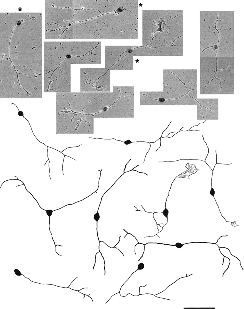
Fig. 10.
Photomicrographs and camera lucida drawings of embryonic day (E)15 trigeminal ganglion (TG) neurons cultured in the presence of neurotrophin-3 (NT-3) for 3 days. These neurons are mostly large cells with short primary neurites and focal arbors. Note the presence of both parvalbumin immunopositive (marked with asterisks) and TrkA immunopositive (unmarked) cells. Scale bar = 100 µm.

