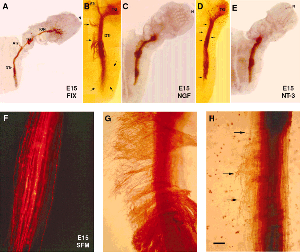
Fig. 2.
Effects of neurotrophins on embryonic day (E)15 central trigeminal axons. Low power photomicrographs of E15 of wholemount cultures of intact whisker pad-trigeminal ganglion (TG)-brainstem (A,C,E) or brainstem with TG (B,D) explants. Trigeminal axons are labeled by inserting 1,1′-dioctadecyl-3,3,3′,3′-tetramethylindocarbocyanine perchlorate (DiI) implants into the ganglion, and the fluorescent label is photoconverted. In control preparation (A), trigeminal pathway is fixed immediately after dissection. At this age, in cultures grown in serum-free culture medium (SFM), central trigeminal axons remain unbranched, and tightly fasciculated, as in normal control cases (F). In the presence of nerve growth factor (NGF; B,C), central trigeminal axons leave the tract, and extend medially or laterally into the brainstem (arrows). In the presence of neurotrophin-3 (NT-3; D,E), these axons emit collaterals and arborize along the trigeminal tract (arrows). Higher magnification views of descending trigeminal tract axons following NGF (G) and NT-3 (H) treatment. Scale bar = 600 µm in A,C,E; 300 µm in B,D; 75 µm in F–H.

