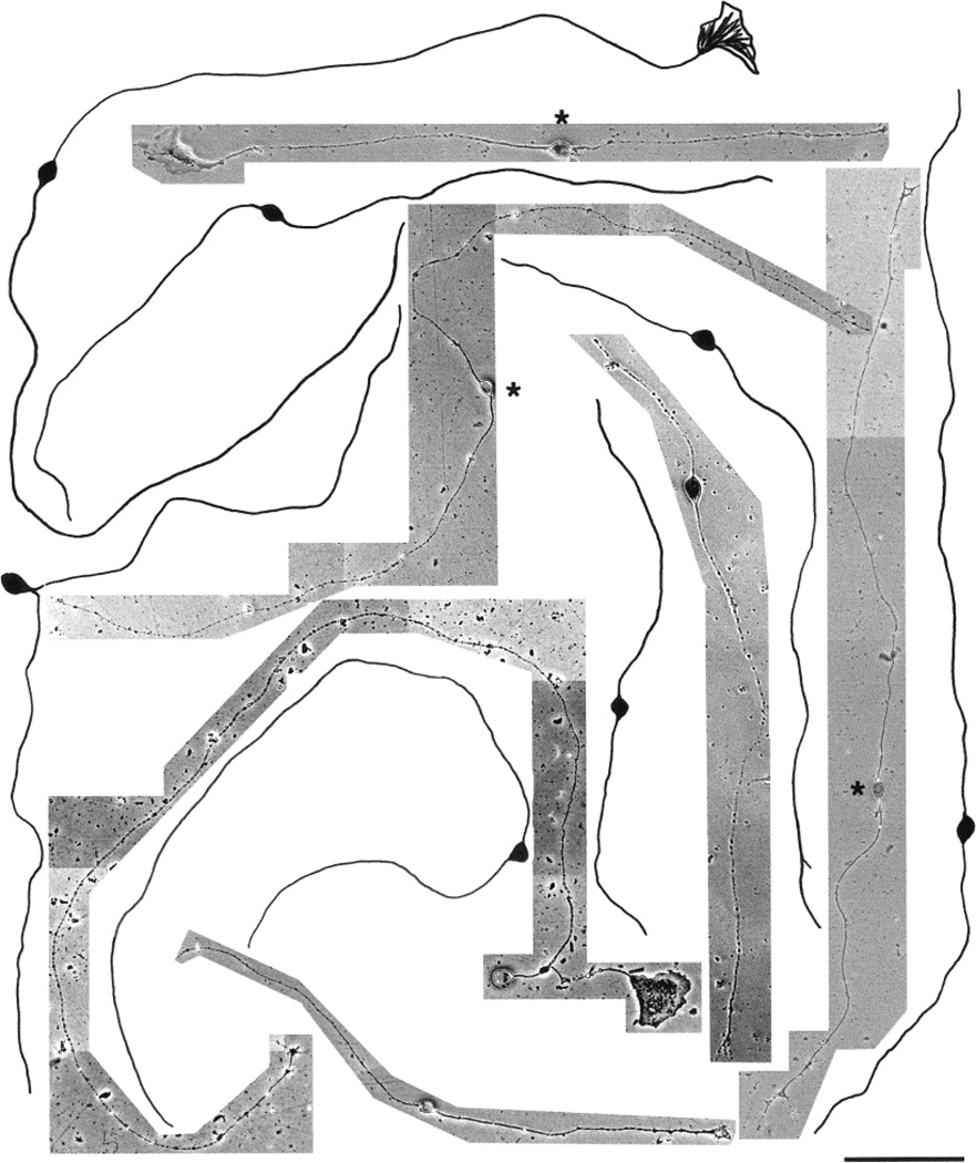
Fig. 9.
Photomicrographs and camera lucida drawings of embryonic day (E)15 trigeminal ganglion (TG) neurons cultured in the presence of nerve growth factor (NGF) for 3 days. These neurons are mostly small cells with very long, unbranched primary neurites. Note the presence of both parvalbumin immunopositive (marked with asterisks) and TrkA immunopositive (unmarked) cells. Scale bar = 100 µm.

