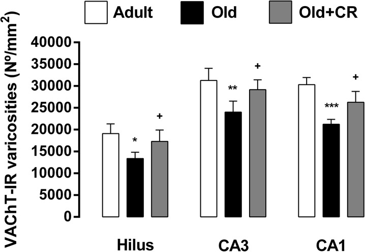
Fig. 6.
Graphic representation of the areal density of VAChT-IR varicosities in the dentate hilus and CA3 and CA1 subfields of 12-month adult control (adult), 24-month-old control (old), and 24-month-old caloric-restricted (old + CR) rats. Note that there was a significant reduction of the density of VAChT-IR varicosities in the dentate hilus (30 %, p < 0.05), CA3 (23 %, p < 0.01), and CA1 (30 %, p < 0.001) subfields of 24-month-old control rats when compared to 12-month adult control rats. Conversely, it was found a significant increase of the density of VAChT-IR varicosities in the dentate hilus (29 %, p < 0.05), CA3 (21 %, p < 0.05), and CA1 (38 %, p < 0.05) subfields of 24-month-old caloric-restricted rats when compared to the 24-month-old control rats. There are no statistically significant differences in the density of VAChT-IR varicosities in the hilus, CA3, and CA1 subfields between 24-month-old caloric-restricted rats and 12-month adult control rats. Data are presented as mean ± SD. *p < 0.05, **p < 0.01, and ***p < 0.001 versus 12-month adult control group; + p < 0.05 versus 24-month old control group

