
Figure 1. Regression of the hyaloid vessels is macrophage-dependent.
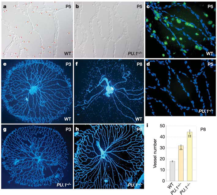
a–d, Hyaloid vessel preparations from wild-type (a, c) and PU.1−/− (b, d) mice at P5. a, b, Differential interference contrast illumination indicating the presence of macrophages in wild-type mice (a, with red dots adjacent) and the absence of macrophages in PU.1−/− mice (b). c, d, Fluorescent immunostaining for macrophages (F4/80, a macrophage-specific marker, green) and nuclei (blue). e–h, Hyaloid vessels from wild-type (e, f) and PU.1−/− (g, h) animals of the indicated ages stained with Hoechst 33258. i, Hyaloid vessel number in wild-type, PU.1−/− and PU.1−/− mice at P8. All error bars are standard errors. Significance levels: three asterisks, 0.0001 < P < 0.001; four asterisks, P < 0.0001. Original magnification: ×50 (e–h); ×400 (a–d). WT, wild type.