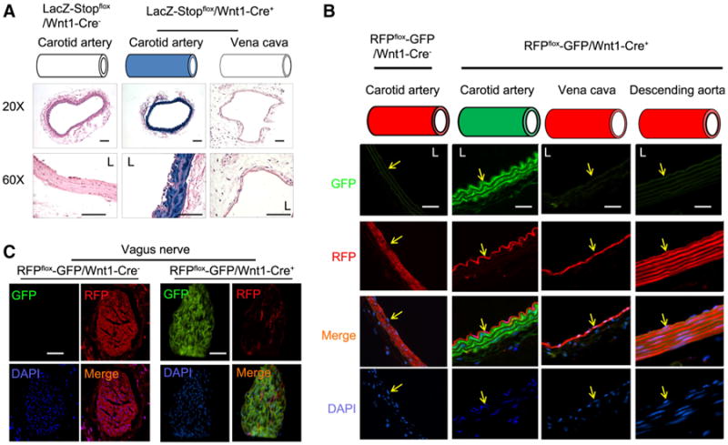
Fig. 1.

Characterization of Wnt1-Cre-mediated expression of reporter genes in vessels. a The common carotid artery and vena cava were collected from LacZ-Stopflox/Wnt1-Cre+ mice or control mice; X-Gal staining was performed. The blue color (X-Gal positive) was only found in the SMCs in the common carotid artery in LacZ-Stopflox/Wnt1-Cre+ mice (n = 5). b, c The cryo section of the common carotid artery, vena cava, and vagus nerve (c) was prepared from RFPflox/flox-GFP/Wnt1-Cre+ or control mice. The RFP and GFP fluorescence was photographed. DAPI was used to counter stain nuclei. Only the SMCs in carotid artery and vagus nerve were positive for GFP. Represented data were from five mice in each group (scales 50 μm, L, lumen; yellow arrows in b indicate the endothelium)
