Fig. 3.

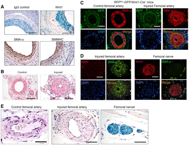
Wnt1 is not expressed in neointima cells in artery injury models. a X-Gal-positive cells did not express Wnt1 in neointima cells from artery grafts created in LacZ-Stopflox/Wnt1-Cre+ mice. The isotype IgG was used as control. Immunostaining of SMC markers, SMA-α and SMMHC, in artery grafts was performed and shown in the lower panel. b Wire injury of the femoral artery induced neointima formation after 1 month (n = 5). c Wnt1-lineage cells are not found in the neointima cells after femoral artery injury. Femoral artery injury was performed in RFPflox-GFP/Wnt1-Cre+ mice. Double staining of GFP and SMA-α was performed in control and injured femoral artery. The right panel shows the injured femoral artery with neointima formation. The non-injured contralateral femoral artery is shown in the left panel (n = 4). d Wnt1 is not expressed in neointima cells in the injured femoral artery. Wnt1 and SMA-α co-immunostaining were performed in the injured femoral artery and normal femoral nerve. Wnt1 expression was only found in femoral nerve. e Femoral artery injury was performed in LacZ-Stopflox/Wnt1-Cre+ mice. X-Gal staining was performed in control and injured femoral arteries (scales in panel a 100 μm; in panel b–e 50 μm; n = 4)
