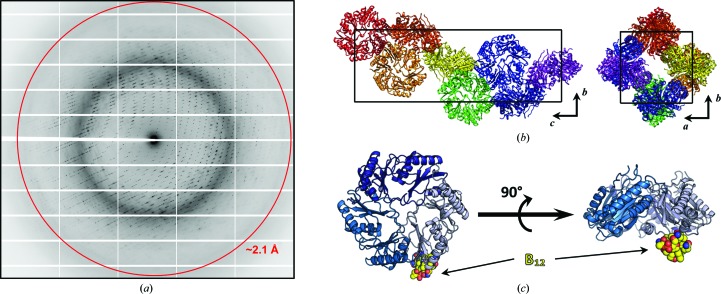
Figure 3.
Overview of cpEutL–B12 structure determination. (a) A representative diffraction image obtained from the X-ray experiment. The red ring represents the 2.1 Å resolution cutoff applied during data reduction. (b) The tetragonal unit cell of the cpEutL crystal contains a single trimer in the asymmetric unit. In the diagrams of the unit cell, each asymmetric unit is colored differently. (c) The structure of cpEutL bound to the hydroxocobalamin ligand (yellow spheres) shows the ligand protruding from the luminal face of the trimer.

