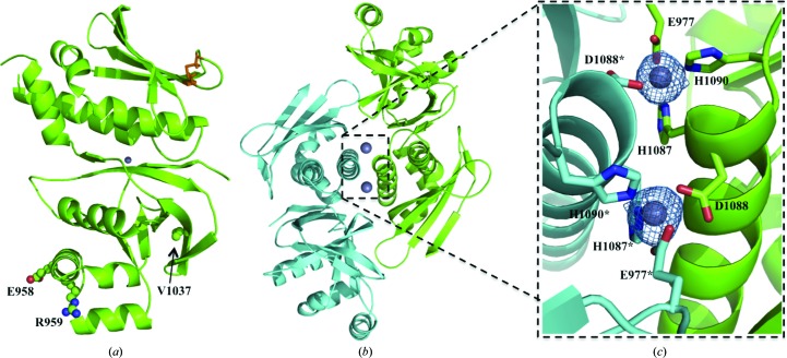
Figure 3.
(a) X-ray crystal structure of PatG-DUFsp. represented as a cartoon. The disulfide bond between Cys1136 and Cys1142 is shown as orange sticks and the Zn2+ ion is shown as a grey sphere. PatG-DUFsp. amino acids which differ from those in PatG-DUFdi are highlighted in ball-and-stick representation for clarity. (b) X-ray crystal structure of the PatG-DUFsp. dimer represented as a cartoon. (c) Enlargement of the Zn2+-coordination site. Glu977, His1087, Asp1088 and His1090 are shown as green sticks and Glu977*, His1087*, Asp1088* and His1090* are shown as cyan sticks. Difference electron density (F o − F c) contoured at 3σ with phases calculated from a model which was refined without Zn2+ present is shown as a blue isomesh.

