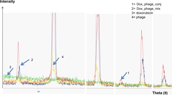
Figure 2.
Magnified spectra of X-ray diffraction from crystallized samples of doxorubicin, phages, and conjugated phages.
Notes: Line 1 (red) represents doxorubicin-conjugated phages, and Line 2 (blue) represents doxorubicin and phage mixture. Line 3 (green) represents doxorubicin, and Line 4 (yellow) represents unconjugated or naked phages. This is the magnified spectra showing the changes in the magnitude of the spectra. An additional peak is evident in conjugated phages, suggesting a change in the structure.
Abbreviations: DOX_phage_conj, doxorubicin conjugated phages; DOX_phage_mix, mixture of doxorubicin and phages.

