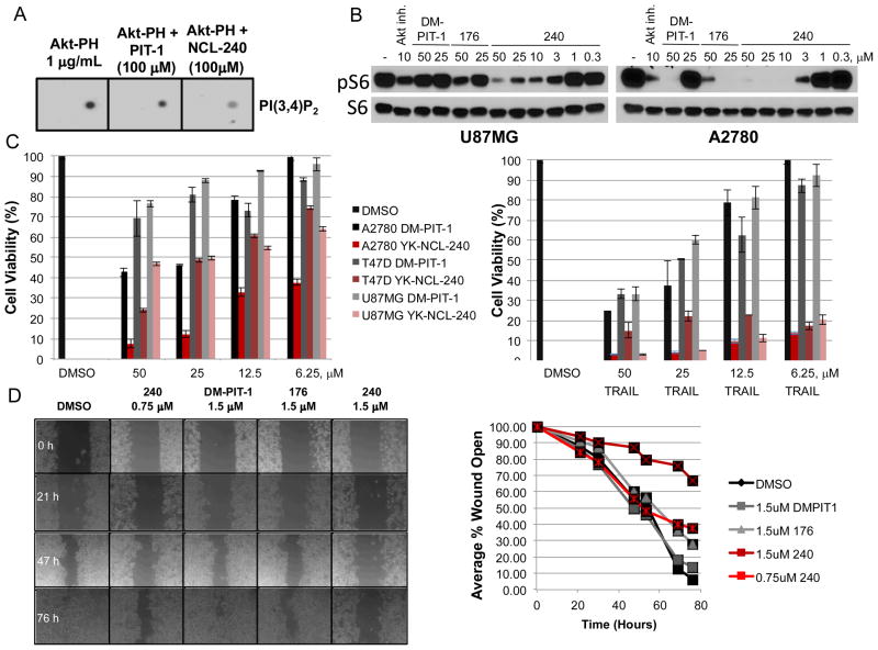
Figure 2.
Increased activity of 1ea. A) Increased inhibition of PI(3,4)P2/Akt-PH domain binding by 1ea.Lipid overlay experiment was performed as performed as described in.3 B) Inhibition of S6 phopshorylation by 1ea. U87MG or A2780 cells were treated with indicated concentrations of inhibitors for 7 hr, followed by Western blotting using phopsho-S6 and total S6 antibodies.C) Increased cytotoxicity of 1ea compared to DM-PIT-1 in multiple cell types. Cells were treated with indicated concentrations of inhibitors alone or in combination with 1 μg/ml TRAIL for 24 hr. Cell viability was determined using CellTiter-Glo assay. D) 1ea efficiently blocks migration of A2780 cells. Wound healing assay was performed as previously described.4 Cell monolayers were treated with indicated concentrations of inhibitors, followed by a scratch wound. Size of the cell free area was photographed at indicated periods of time. Quantification of open wound area is shown on the right.

