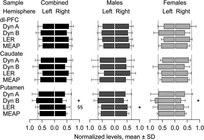
Figure 3.
Comparison of the opioid peptide levels between the left and the right hemispheres of the dl-PFC, caudate, and putamen. Data are shown as means ± SD of the normalized levels in the LH [L/(L + R)] and RH [R/(L + R)]. Dashed lines at a 0.66 value mark 2-fold differences between hemispheres. Significance levels in ANOVAs followed by ANCOVAs when appropriate: Lateralization effects, *P < 0.05; lateralization × sex interactions, §§p < 0.01.

