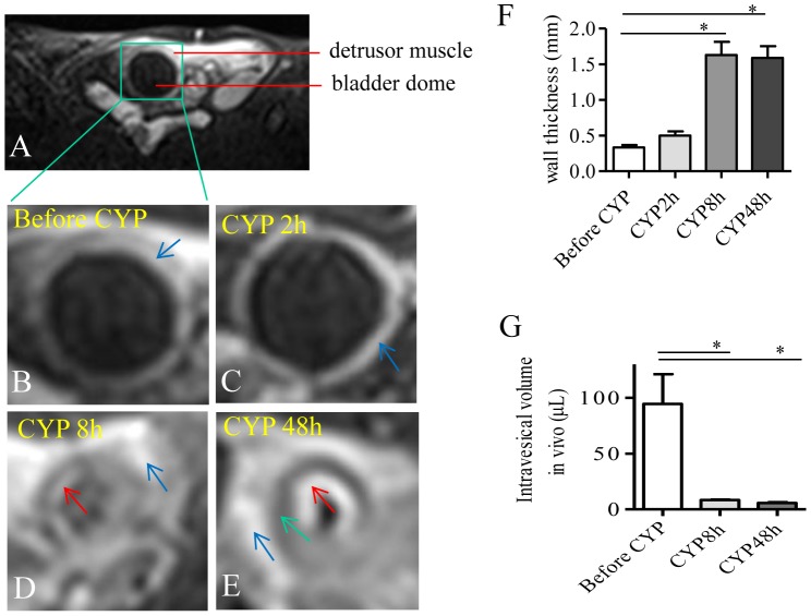
Figure 1. MRI visualization demonstrated an increase in the thickness of bladder wall and a decrease in the volume of bladder dome during CYP-induced cystitis.
A spin-echo T2-weighted MR sequence was performed to visualize the urinary bladder in the pelvis on axial sections (A). The same animal was scanned before (B) and after CYP treatment (C-E). At 2 h after cystitis was induced, the anatomy of the urinary bladder appeared similar to control. At 8 h and 48 h post cystitis induction, the thickness of the bladder wall was significantly increased (F) which was accompanied with a decrease in intravesical volume (G). Summary results were from 5 animals before and after CYP injection. *, p<0.05.

