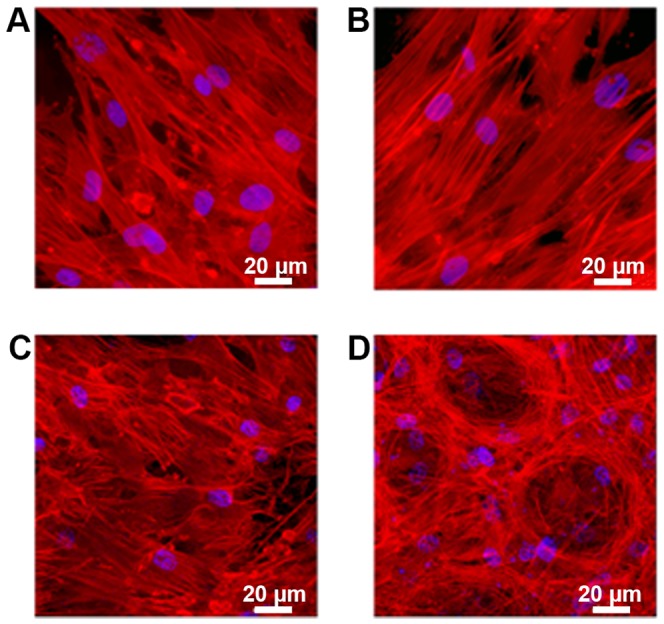
Figure 5. Cellular attachment observed by fluorescence microscopy.

(A and B) human mesenchymal stem cells(MSCs) and (C and D) MSCs cultured in osteogenic differentiation media (MSC-OM) with (A and C) plain PMMA bone cement and (B and D) PMMA bone cement loaded with gentamicin and AgNP incubated for 7 days (MSC) and 21 days (MSC-OM), respectively.
