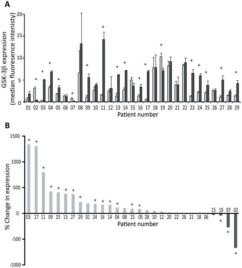
Figure 1. Expression of GSK3 in NSCLC tumour tissue in comparison to patient-matched normal lung tissue.
Three distinct samples from patient-matched normal (N1-3) and tumour (T1-3) tissues were analysed using Luminex (xMAP) technology to determine the level of GSK3β expression. A: Quantified data for all 29 patients. Each bar represents the average expression for normal (N1-3; light grey) or tumour (T1-3; black) tissue for each patient (mean ± SEM). The strength of evidence for a difference in expression between the normal and tumour samples was determined by a Kruskal-Wallis test, and * indicates p<0.05. B: The percentage change in GSK3 expression in tumour samples in comparison to patient-matched normal tissue where patients are ranked in order of the extent of the percentage change in expression.

