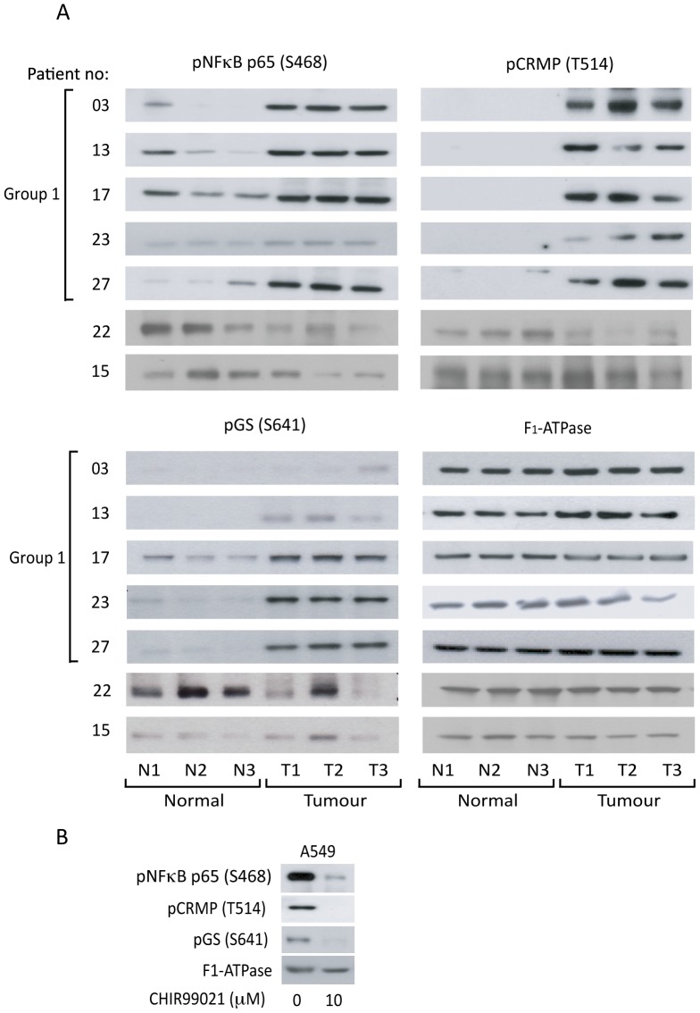
Figure 5. Phosphorylation of downstream GSK3 substrates in selected patients.
A: Three distinct samples from normal (N1-3) and patient-matched tumour (T1-3) tissues were separated on SDS-PAGE gels. Expression of GSK3 and phosphorylation of GS (S641), NFκB p65 (S468) and CRMP (T514) was determined by western blotting with specific antibodies as indicated. An anti-F1-ATPase antibody was used as a control for protein loading. B: A549 cells were cultured in the absence and presence of 10 µM CHIR99021. Protein was extracted and lysates were separated on SDS-PAGE gels. Expression of GSK3 and phosphorylation of GS (S641), NFκB p65 (S468) and CRMP (T514) was determined by western blotting.

