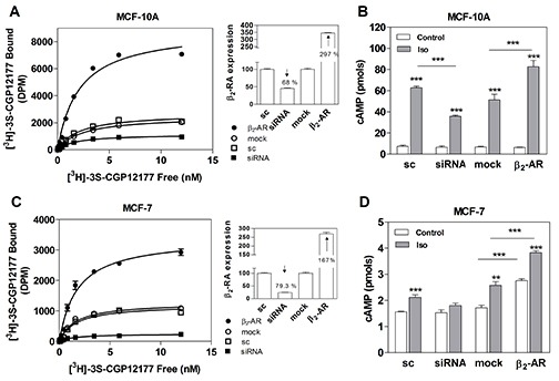
Figure 4. β2-AR overexpression and knock-down in MCF-10A and MCF-7 cells.

(A) Quantification of β2-AR in MCF-10A and (C) MCF-7 cells transfected with scrambled siRNA (sc), β2-AR-targeted pooled siRNA (siRNA), pcDNA3.1 (mock) or the plasmid codifying for the β2-AR. Panels A and C depict the saturation analysis performed with the β-AR radioligand [3H]-GCP 12177. The results are expressed as the percentage of the scrambled or the mock in whole cells at 4 °C. The modification of the expression of β-AR in the cells is shown in insets as a percentage of the sc or mock. (B) Total cAMP production in MCF-10A cells or (D) MCF-7 cells transfected with sc, siRNA, mock or β2-AR and incubated or not (control) with 1 μM Isoproterenol (Iso). Data represent the mean ± s.e.m. of two independent experiments. Statistical significance was assessed using ANOVA followed by a Bonferroni test. *** p<0.001.
