
Figure 4. TRIM16 protein expression is reduced in metastatic melanoma and correlates with overall survival risk in stage III disease.
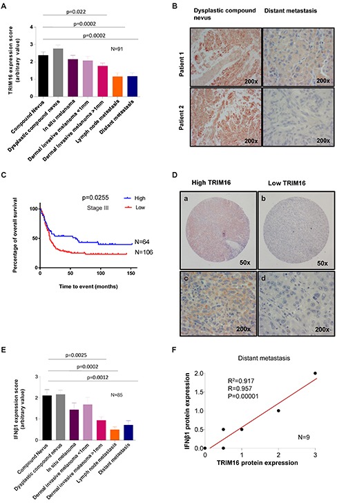
(A) The level of TRIM16 protein expression was analysed by immunohistochemistry by two observers blinded to clinical outcome using a polyclonal TRIM16 antibody and graded on an arbitrary scale of 0-4. The score of TRIM16 expression was collected from 91 patient samples with 13 samples per tumor type/ melanoma stage. The statistical comparisons were performed using student's t-test. A statistically significant difference for TRIM16 expression level between melanoma and compound nevi is indicated by p value. (B) Representative immunohistochemistry images of TRIM16 staining in two samples of dysplastic compound nevi and distant metastases. (C) Kaplan-Meier curve of overall patient survival for TRIM16 protein expression level, using the median value to subdivide the cohort for ‘high’ and ‘low’ staining. Patient median survival was 59 months for TRIM16 expression level of >1, and 16 months for TRIM16 expression level of <1. The data was analysed with the Log-rank test. (D) Representative immunohistochemical staining of lymph node metastases showing lymph node metastasis with high (a, c) or low (b, d) TRIM16 expression. (E) The level of IFNβ1 expression in primary and metastatic melanocytic tumors was measured for 85 melanoma patients using immunohistochemical grading after staining with an anti-IFNβ1 antibody. There were 13 samples per tumor type/stage for compound nevus to dermal invasive melanoma >1 mm and 11 samples for lymph node metastasis and 9 for distant metastasis. The statistical analysis was performed by the student's t-test. A statistically significant difference is indicated by **p<0.01 or ***p<0.001, when expression in melanoma was compared with compound nevi. (F) A correlation analysis between IFNβ1 and TRIM16 expression in distant melanoma metastases (n = 9).