
Figure 1. Overexpression of Gα12 in oral squamous cell carcinoma (OSCC) tissues and cell lines.
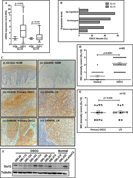
(A) qPCR analysis indicated that Gα12 was significantly overexpressed at the mRNA level in OSCC tissues. As indicated by the box and whiskers plot, OSCC tissues had a median of 2.35-fold increase in Gα12 expression, with a maximum level of 23-fold overexpression compared to normal oral mucosa (NOM). Meanwhile, the median of Gα13 expression is 1.1-fold, suggesting no differential expression between OSCC and NOM. (B) Gα12 mRNA levels were found to be overexpressed in 55.3% of OSCC tissues examined, and majority of the OSCC tissues had down-regulation of Gα13, when using a cut-off fold-change value of ≥ 2. (C) IHC images showing NOM tissues have negative staining for Gα12 (panel i & ii), but the primary tumors and the LN of matched cases showed overexpression of Gα12 (panel iii & iv; panel v & vi). Images were captured using 200x objective. (D) IHC staining analysis indicated that Gα12 is overexpressed in the primary tumors compared to NOM tissues. (E) Expression of Gα12 was also retained in the OSCC cells that metastasized to the lymph nodes (LN). Each symbol represents one patient. Error bars in IHC analyses represent standard error of the means (SEM) of Gα12 expression for the total number of tissues examined. (F) Western blot indicated Gα12 is overexpressed in all OSCC cell lines and absent in normal oral keratinocyte primary cultures. α-tubulin was used as an endogenous control and squamous non-oral HeLa cells transformed to overexpress Gα12 was used as a positive control.