
Figure 4. G12-RhoA signaling axis is involved in OSCC metastasis to cervical lymph nodes (LN).
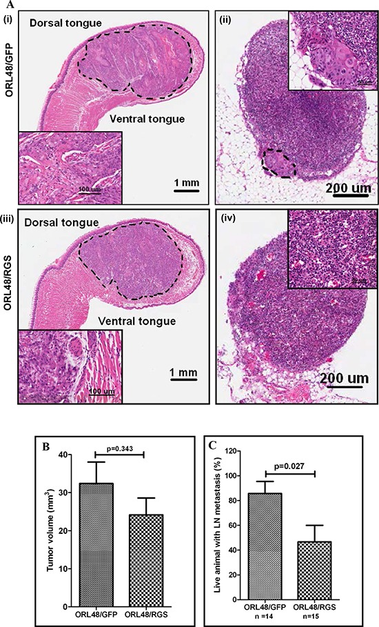
(A) H&E-stained tissue section of the orthotopically implanted OSCC cells into the tongue. ORL48/GFP control cells (i) and ORL48/RGS (iii) formed primary tumors growing into the anterior half of the tongue after day 20 post-implantation. Insets are higher magnification of the tumor area. Histologic evaluation of H&E-stained sections of the representative cervical lymph node showing the metastatic growth of ORL48/GFP control cells in the area rounded by dotted line. Inset is the higher magnification of the metastatic area (ii). Histological section of a non-invaded lymph node in the ORL48/RGS group (iv). (B) The growth of ORL48/GFP (control) primary tumors on the tongue were larger than those of the ORL48/RGS tumors, but the differences were not statistically significant. (C) Increased number of metastatic LN was found in mice carrying orthotopic ORL48/GFP control tumors, as compared to ORL48/RGS tumors. Animals that died of the disease before day 20 post-implantation were not included in the analysis as their LN were difficult to retrieve. The bar chart represents the average percentage of total animals that showed LN metastasis in 3 sets of experiments, while the error bars are SEM for these experiments.