
Figure 2. SapC-DOPS induces lysosomal cell death via membrane permeability and organelle dysfunction.
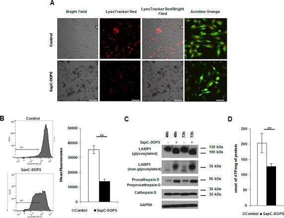
(A) Lysosomes were analyzed by fluorescent microscopy in U251 GBM cells treated with SapC-DOPS (45μM) using LTR or AO at 48h (B) or by FACS analysis using LTR. Numbers indicate the percentage of cells with decreased fluorescence. (C) Lysosomal proteins were analyzed via immunoblot from U251 GBM cells treated with SapC-DOPS (45μM). (D) ATP was measured in U251 GBM cells 48h following treatment with SapC-DOPS (45μM). Error bars show mean + SD, **P<.01. Scale bar 100μm.