
Figure 3. Reduction of IgG impaired LPS-initiated TLR4 signaling pathways in cervical cancer cells.
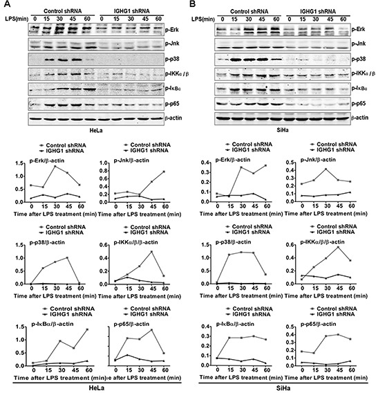
(A) Immunoblot analysis of phosphorylated (p-) signaling molecules (p-ERK, p-JNk, p-p38, p-IKKα/β, p-IκB α, and p-p65) in lysates of HeLa cells stably expressing IGHG1 shRNA or control shRNA stimulated for 0–60min (above lanes) with 100 ng/ml LPS. β-acin was derived from the same samples and used as an internal control. Similar experiments were performed using SiHa cells treated with IGHG1 shRNA or control shRNA (B). The shown results are representative of three independent experiments (upper panel). Phosphorylation levels of the above proteins were quantitated by band density scanning and shown in the lower panel. The values were normalized to the β-actin signal.