
Figure 6. Reduction of IgG downregulated TLR4 expression.
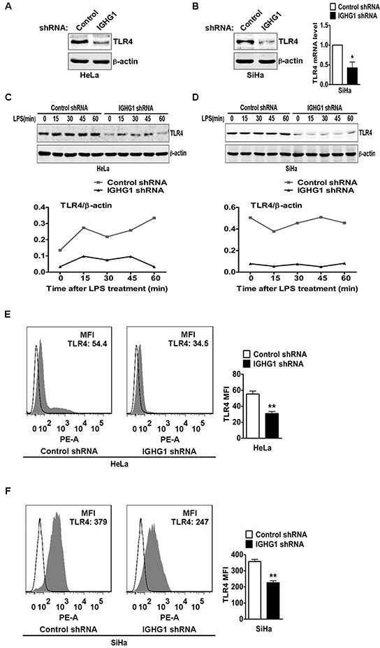
TLR4 expression at protein level in HeLa cells stably expressing IGHG1 shRNA or control shRNA. (A) and IGHG1 silenced SiHa cells or control cells. (B) were measured with immunoblot using anti-TLR4 Antibody. β-actin was used as a quantitative control. TLR4 expression at mRNA level in the above SiHa cells was also measured with RT-qPCR (B). (C) HeLa cells stably expressing IGHG1 shRNA or control shRNA and IGHG1 silenced SiHa cells or control cells (D) were treated with 100 ng/ml LPS. Dynamic changes of TLR4 expression (0, 15, 30, 45, and 60 minutes) were examined with immunoblot using TLR4 antibody. β-actin was used as quantitative control. The results shown are representative of three independent experiments (upper panel). The quantified results of TLR4 expression were shown in the lower panel. The values were normalized to the β-actin signal. TLR4 expressions on plasma membrane in HeLa cells stably expressing IGHG1 shRNA or control shRNA (E) and IGHG1-silenced or control vector-silenced SiHa cells (F) were detected with phycoerythrin-conjugated TLR4 antibody by flow cytometry. The mean fluorescence intensity (MFI) was calculated and shown at the right panel. The data are expressed as mean±S.D. from three independent experiments (*P<0.05; **P<0.01).