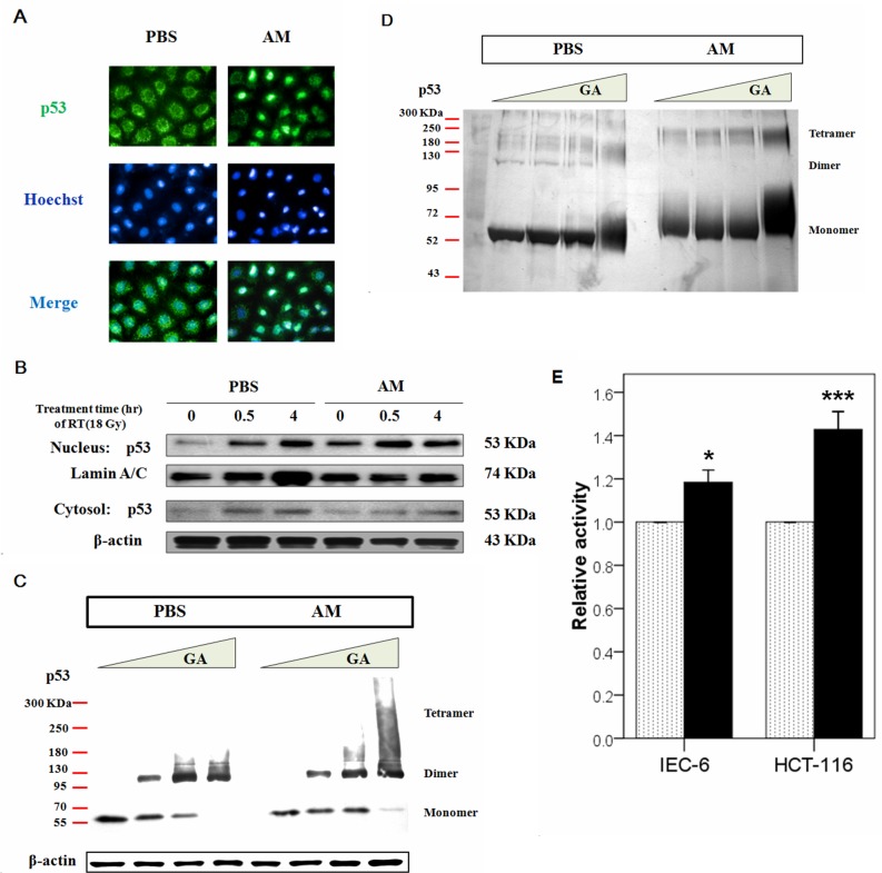
Figure 6. AM increases the nuclear but not cytosolic fraction of p53.
(A) Immunofluorescent staining for p53 was performed in IEC-6 cells that had been treated with AM (4 mM) or vehicle (PBS) for 60 minutes, after which these compounds were washed out. Green color indicates p53 expression, and blue color indicates Hoechst nuclear staining. (B) Analysis of the nuclear and cytosolic fractions of p53 was performed between AM- and PBS-treated cells before and after irradiation. The internal control was lamin A/C for the Western blot assays. (C) Oligomerization assays were performed using glutaraldehyde (GA) for crosslinking. The relative amounts of p53 monomers, dimers, and tetramers, which exhibit different molecular weights, were determined using Western blotting. (D) An oligomerization assay in a cell-free system was performed. Purified human p53 (500 ng) (lanes 1-8) was pretreated with AM (lanes 5-8) for 60 minutes at 37°C and then incubated with 0% (lanes 1 and 5), 0.008% (lanes 2 and 6), 0.016% (lanes 3 and 7), or 0.09% (lanes 4 and 8) GA for 15 minutes at 37°C. Samples were analyzed by SDS/4-20% PAGE, followed by silver staining. (E) The transcriptional activity of p53 was compared between AM (black bar)- and vehicle-treated cells in IEC-6 and HCT 116 cells. Error bars represent the standard error of the mean. * p < 0.05, *** p <0.001.

