
Figure 2. Ligand-dependent PR downregulation.
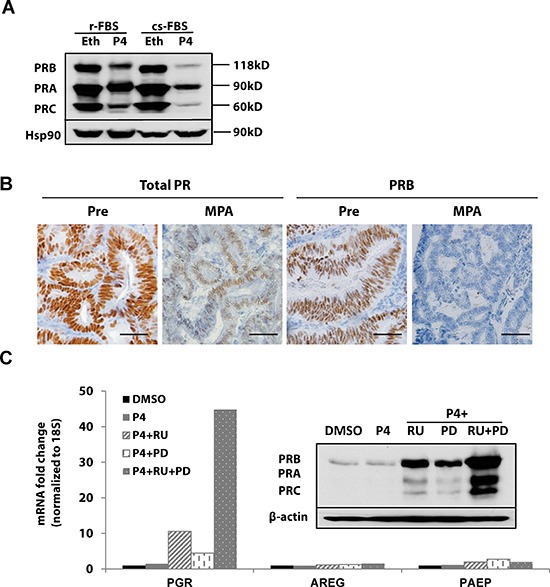
(A) Western blotting: T47D breast cancer cells were grown in DMEM media supplemented with regular fetal bovine serum (r-FBS) or charcoal-stripped serum (cs-FBS), followed by treatment with ethanol (vehicle control) or 100 nM progesterone (P4) for 24h. PR protein was detected using specific PR antibodies, and HSP90 serves as a loading control. (B) Immunohistochemistry: endometrial tumor specimens were collected before or 21 days after MPA treatment (400 mg, intramuscularly) and total PR and PRB expression was assessed by immunohistochemistry. Scale bar = 50 μm. (C) q-PCR and Western blotting: ECC1 cells were treated with DMSO (vehicle control), 100 nM P4, 100 nM P4 and 1μM PR antagonist RU486 (RU), 100 nM P4+ 1 μM MAPK inhibitor PD0325901(PD) or the combination of P4+RU+PD for 24h. mRNA expression of PGR, AREG and PAEP was measured by q-PCR, normalized to 18S, and data displayed as fold-change relative to DMSO control. Comparisons of normalized expression values (ΔCt) employed the conventional ΔΔCt fold change method. The insert is PR protein expression after the same treatment; β-actin, loading control.