
Figure 4. Histone deacetylase (HDAC) inhibition restores PR mRNA and protein expression in Type I Ishikawa H cells.
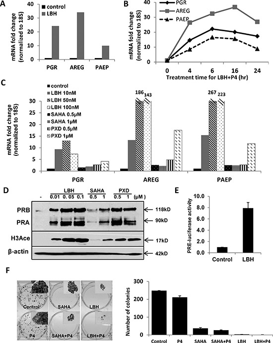
(A) q-PCR analysis. Ishikawa H cells were treated with DMSO control or 20 nM LBH589 (LBH) for 24 h. PGR, AREG and PAEP mRNA expression was normalized to 18S, and all q-PCR data are displayed as fold-change relative to DMSO control. Comparisons of normalized expression values (ΔCt) employed the conventional ΔΔCt fold change method. (B) PR expression and activity corresponding to time course of P4 stimulation: Ishikawa H cells were treated with 20 nM LBH +100 nM P4 for the indicated times, and PGR, AREG and PAEP mRNA expression was quantified by q-PCR and normalized to 18S. (C) q-PCR analysis. Ishikawa H cells were treated with three different HDAC inhibitors (LBH589, SAHA and PXD101) at the indicated concentrations for 24h. PGR, AREG and PAEP mRNA expression was quantified by q-PCR and normalized to 18S. (D) Western blotting. Expression of PR protein in Ishikawa H was measured after treating with the three HDAC inhibitors. The presence of histone H3 acetylation indicates drug effect, and β-actin serves as a loading control. (E) PRE-luciferase assay. Ishikawa H cells were treated with 20 nM LBH589 for 24h and studied using a PRE-luciferase assay. The PRE-luciferase activity was normalized to total protein concentration. (F) Colony formation assay. Ishikawa H cells were treated in the presence or absence of HDACi for 2 weeks, and resulting colonies were stained with crystal violet (left panel, insets are 5X) and the number of colonies recorded (right panel). Error bar, SD.