
Figure 6. Impact of miRNAs on PR expression in endometrial tumors and cells.
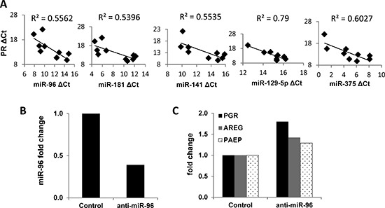
(A) Correlation of miRNA expression with PGR expression in matched non-malignant endometrial tissue vs. endometrioid adenocarcinoma (n=5). mRNA expression of PGR was measured by q-PCR and normalized to 18S and miRNA expression was measured by q-PCR and normalized to RNU48, and both data are displayed as ΔCt value relative to the DMSO control. (B) miR-96 was decreased by transfecting the miR-96 inhibitor into Ishikawa H cells. (C) PR expression was restored by inhibiting miR-96 in Ishikawa H cells. Comparisons of normalized miRNA expression values (ΔCt) employed the conventional ΔΔCt fold change method.