
Figure 7. Systematic analysis of strategies to restore functional PR expression in distinct models of endometrial cancer.
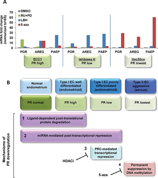
(A) Three endometrial cancer model cell lines, ECC1, Ishikawa H and Hec50co cells were treated with DMSO control, 1 μM RU486+1 μM PD0325901 for 24h, 20 nM LBH589 for 24h or 100 nM 5-aza for 3 days. mRNA expression of PGR, AREG and PAEP were measured by q-PCR, normalized to 18S and displayed as fold change to DMSO control. Comparisons of normalized expression values (ΔCt) employed the conventional ΔΔCt fold change method. In ECC1 cells, which express PR at baseline, treatment with the progestin antagonist RU486 and the MAPK inhibitor PD0325901 validates the dynamic PR regulation by ligand-mediated degradation. In Type I Ishikawa cells, only an HDACi effectively restores functional PR, whereas a hypomethylating agent is necessary for Type II Hec50co cells. (B) Proposed model of PR downregulation mechanisms during endometrial cancer progression.