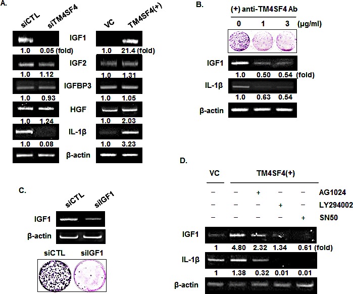
Figure 5. IGF1R activation of TM4SF4-overexpressing A549 cells is induced by enhanced expression of IGF1 via NF-kappaB activation.

(A) RT-PCR analysis of components of IGF1R signaling pathway and IL1 beta in TM4SF4- knockdown or overexpressing A549 cells. HGF served as negative control and beta-actin as internal control. Band intensities were measured using Image J software, normalized to β-actin and fold increase were indicated. (B) Colony formation assay and RT-PCR analysis of effect of blocking of TM4SF4 by anti-TM4SF4 antibody in TM4SF4-overexpressing A549 cells. (C) Colony formation assay of IGF1-knockdown A549 cells. (D) Effects of IGF1R, PI3K, or NF-kappaB inhibition on the expression of IGF1 and IL1 beta. TM4SF4-overexpressing A549 cells were treated with AG1024 (10 μM for 24 h), LY294002 (50 μM for 48 h) and SN50 (20 μM for 24 h) and analyzed by RT-PCR.
